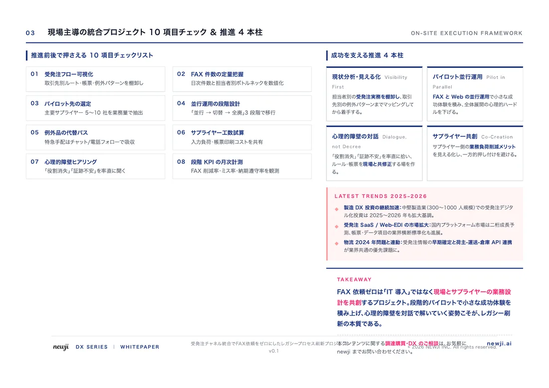
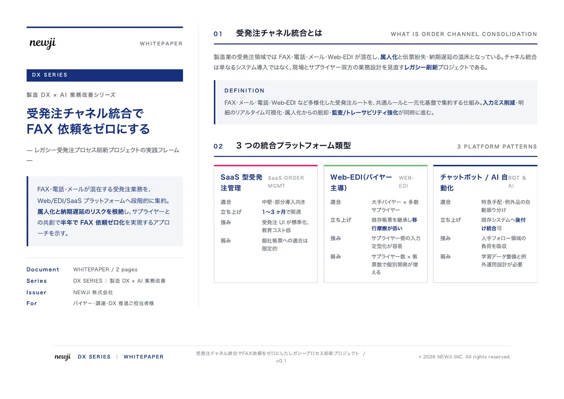
- お役立ち記事
- Polymer blend domain analysis using atomic force microscope (AFM) phase images
スタートアップから大手まで。
調達・受発注をAIで標準化。
相見積比較も進捗管理もAIが下支え。取引先は招待で完全無料。

Polymer blend domain analysis using atomic force microscope (AFM) phase images

When it comes to analyzing polymer blends, scientists often utilize advanced techniques to ensure precise observations and results. One such technique that has gained significant attention is the use of an atomic force microscope (AFM) to study polymer blend domains. AFM provides detailed images of the material’s surface, and its phase images, in particular, offer important insights into the composition and distribution of different polymer components. In this article, we’ll explore how AFM phase images are applied in the analysis of polymer blend domains.
目次
Understanding Polymer Blends
Polymer blends are combinations of two or more polymers mixed to create a material with desirable properties specific to particular applications. These materials can be engineered to exhibit improvements in qualities such as toughness, flexibility, or resistance to various environmental factors. Analyzing these blends is crucial because the performance of a polymer blend is highly dependent on the distribution and morphology of its constituent polymers.
The Role of AFM in Polymer Studies
An atomic force microscope is a powerful tool in material science, renowned for its ability to provide high-resolution images of surfaces. Unlike other microscopy techniques that rely on lenses and light, AFM uses a delicate cantilever with a sharp probe at its tip to scan the surface of a sample. When the probe interacts with the surface, it experiences forces that cause the cantilever to deflect. These deflections are recorded and used to generate detailed topographical maps of the surface at the nanoscale.
AFM is particularly effective for studying polymer blends because it can reveal the surface morphology and phase distribution without damaging the sample. This makes it ideal for studying the nanoscale phase separation that often occurs in polymer blends.
Phase Imaging in AFM
Phase imaging is a specific mode of AFM that analyzes variations in the material’s mechanical properties at the surface. While the ordinary topographical image gives information about the physical surface structure, the phase image provides insights into the material’s compositional or mechanical differences. This is key when studying polymer blends, as the phase image can distinguish between domains composed of different polymer components based on variations in stiffness, adhesion, or viscoelastic properties.
Analyzing Polymer Blend Domains
When using AFM phase images for polymer blend analysis, researchers are often interested in understanding the size, shape, and distribution of different polymer regions within the blend. These domains can significantly influence the material’s overall properties.
Identifying Domain Structures
AFM phase images allow scientists to visually identify distinct domain structures within a polymer blend. Differently shaded regions in the phase image represent areas with varying mechanical properties, suggesting the presence of different polymer types. This enables researchers to ascertain whether the polymers are well-mixed, or if there are distinct phase-separated regions, which can impact mechanical strength or flexibility.
Quantitative Measurement of Domains
Beyond visual identification, advanced image processing software can be used to quantitatively measure the size and distribution of these domains. Statistical analysis of AFM images can provide data on average domain size, distribution patterns, and the extent of interfacial interactions between different polymer types. This is critical for correlating the blend’s microstructural characteristics with its macroscopic properties.
The Importance of AFM Phase Imaging
AFM phase imaging serves as a non-invasive and highly effective method for the detailed study of polymer blends at the nanoscale. By providing insights into the phase separation and distribution of components within the blend, this technique assists in tailoring materials with specific desired properties for applications ranging from flexible electronics to robust construction materials.
Applications and Future Directions
With ongoing advancements in AFM technology, researchers are developing even more sensitive and accurate instruments that can reveal ever smaller and more complex structures within polymer blends. As these technologies evolve, the potential for creating even more advanced materials grows, opening new frontiers in industries such as bioengineering, coatings, and packaging.
In the future, ongoing improvements to AFM and phase imaging will likely enhance the ability of scientists to investigate blends under different conditions, such as varying temperature or stress. This information can provide further guidance in developing materials that are optimized for performance under specific operational situations.
To sum up, atomic force microscopy and its phase imaging capabilities offer invaluable tools for the analysis and optimization of polymer blend domains. By understanding and controlling the microstructure of these complex materials, researchers and manufacturers can drive innovation and create products with enhanced performance and reliability.
この記事の理解を深める
無料ホワイトペーパーをプレゼント
製造業の現場で使える実務資料(PDF)を無料でお届けします。"こんな資料が届きます" ↓ 下のボタンからどうぞ。
PRODUCT — 製造業向け 調達・受発注クラウド
この記事の課題、
newji で解決しませんか?
newji は、製造業の調達・受発注に特化したクラウド/AIエージェント。見積依頼・発注書作成・進捗管理・承認をひとつの画面に集約し、AIが比較と異常検知を担当。最後の「GO」だけ人が押す仕組みです。
- 見積〜発注〜納期を一元管理。催促・転記のムダをゼロに
- AIが相見積もり比較と異常検知。あなたは判断だけに集中
- 取引先は「招待」で完全無料。自社コストだけで取引先ごとデジタル化
※ 取引先から招待された企業様は完全無料でご利用いただけます